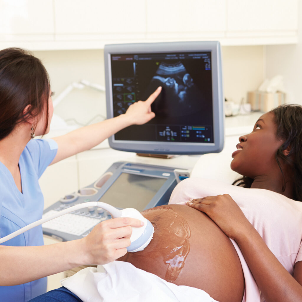
Ultrasounds at A.T. Radiology
Also known as sonography, ultrasound uses high-frequency sound waves to generate images of the body’s internal anatomy. Just like with sonar used by submarines, as the sound waves bounce off an object, they create echoes. In the case of ultrasound imaging, the echoes created when sound waves bounce off internal organs and tissues are translated by a computer into images on a screen. Doctors then use these images to diagnose abnormalities within the body. In the case of prenatal imaging, ultrasound can help ensure that a baby is developing normally.
Ultrasound testing does not use any form of radiation, so it is widely used where X-ray-based tests are not, such as when evaluating the body’s reproductive system. A modified ultrasound technique—called Doppler—is used to capture moving images of the heart and blood vessels.